

Revolutionize Your Imaging of Soft Tissue Structure
Internal Structure Analysis without Complex Sample PreparationX-ray imaging provides a unique opportunity for the exploration of low-density biological specimens such as 3D cultures, whole organs, tumors and embryos. The non-destructive nature of the approach means your sample remains intact for subsequent evaluations using different microscopy techniques or further analyses. The structural information obtained with X-ray imaging complements the functional or specific localization information found with fluorescence microscopy and bridges the resolution gap to the ultrastructural information captured using electron microscopy.
Sample courtesy of the University of Radboud, Netherlands
Capture Structural Information across Length Scale


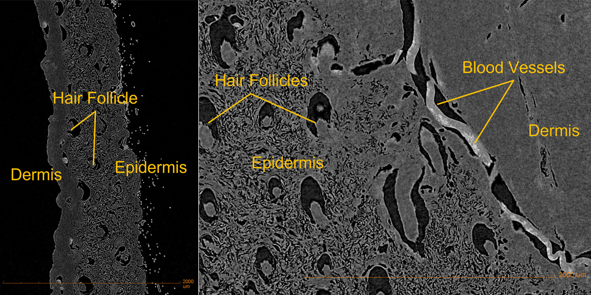

Single 2D projections from a high-resolution 3D scan of a piece of dissected mouse skin imaged with ZEISS Xradia Versa
Single 2D projections from a high-resolution 3D scan of a piece of dissected mouse skin imaged with ZEISS Xradia Versa
Gain Structural Insights Down to the Nanoscale
High-resolution acquisition is often vital for visualization of structures of interest in tissue specimens. Using non-destructive X-ray microscopy, key components of tissues such as skin can be clearly visualized, including blood vessels and hair follicles. The two-stage magnification of ZEISS Xradia Versa enables the capture of high-resolution insights without having to cut the sample into smaller pieces.
Optimal Contrast in Low Density Specimens
High Contrast in Soft Tissues
Visualizing internal structures can be challenging in soft tissue specimens due to samples having only small differences in X-ray absorption. An instrument with high contrast capability is therefore essential for optimal image quality. The optimized objective lenses of the ZEISS Xradia Versa play a key role in ensuring the best possible image quality, even when differences in absorption contrast are very low in your soft tissue specimens.


Unstained mouse embryo imaged with ZEISS Xradia Versa with high contrast capability.
Unstained mouse embryo imaged with ZEISS Xradia Versa with high contrast capability. Courtesy of Dr. Yukako Yagi, Massachusetts General Hospital, USA
Image Unstained Specimens
Sometimes it is desirable to image soft tissue specimens, such as embryos, without any staining agents. For unstained samples, the superior contrast capability of ZEISS Xradia Versa provides an excellent means to still visualize internal structures using absorption contrast.
Capture Interfaces Using Propagation Phase Contrast
If staining is not possible, but differences exist in X-ray refractive index (for example membranes or cell walls), alternative contrast methods such as propagation phase contrast are available with ZEISS Xradia Versa. Propagation phase contrast highlights the interface between components of the specimen with different X-ray refractive indices and therefore specimen structure can be visualized even without staining.
Gain Vast Volumes of Internal Information
Compare Models or Treatment Groups
When natural differences in X-ray absorption are insufficient for visualization of the structures of interest in the tissue, contrast enhancing agents can accentuate the differences. Many different staining approaches can be used2 and, for samples such as embryos, this can generate a huge amount of information when imaged at high resolution using the X-ray microscope. This imaging approach affords great opportunities for comparative studies between different genetic models or disease and treatment groups.
Improve Signal-To-Noise Ratio and Throughput of 3D Acquisitions


Mouse lung tissue. Equivalent single 2D sections through reconstructed datasets acquired with the same parameters (3001 projection images). Left: standard FDK reconstruction. Right: Deep learning reconstruction (DeepRecon).
Simultaneous Reduction of Noise and Acquisition Time with Deep Learning Reconstruction
For soft tissue specimens with low density and small differences in contrast, reducing the noise in the reconstructed images can make a significant difference to the structures that can be visualized. Deep Learning reconstruction not only increases the signal-to-noise ratio of the 3D reconstructed datasets, but it also increases throughput as fewer 2D projections are required. ZEISS DeepRecon provides a straightforward workflow for deep learning reconstruction and can uncover small details and structures in low density soft tissue specimens that are otherwise hidden by noise.


Related Products


-
1
Q Chu et al. (2020), https://onlinelibrary.wiley.com/doi/10.1002/advs.201903592
-
2
B.D. Metscher (2009), https://bmcphysiol.biomedcentral.com/articles/10.1186/1472-6793-9-11