ZEISS EVO Solutions for Life Sciences

ZEISS EVO Solutions for Life Sciences
The scientific study of life science is pertinent to improving life, from improving health to saving the environment and growing better crops. Our understanding of life science depends on the characterization of biological specimens from macro down to nanoscale details. Combining powerful scanning electron microscopy with analytical solutions and dedicated software creates excellent images and extracts meaningful information from your microscopy images to answer your scientific questions.
The Scanning Electron Microscope (SEM) is now an essential tool for researchers. However, choosing the correct configuration and capability and training users of all skill and experience levels has always been tricky. Multiple-use instruments require flexibility, analytical capability, excellent productivity, reliability and stability. Finding the region of interest has to be fast, and the images you can create must be exceptional to understand the smallest features and gain unique insights into your biological sample.
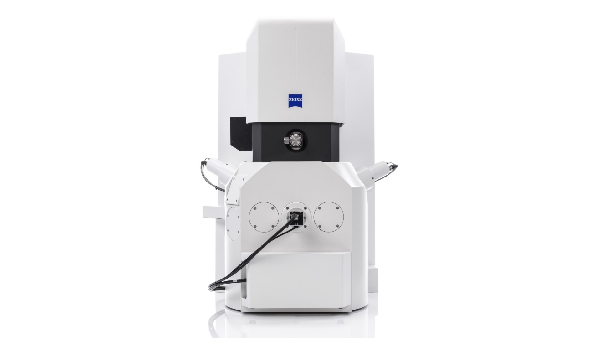
ZEISS EVO is the life science researchers’ stable, intuitive, uncompromising SEM for research applications. It is easy to buy, easy to train and easy to use. In addition, we have created “Solution Bundles” for life science applications designed for customer workflows. They combine our comprehensive ZEN software ecosystem, with its suite of automation and image analysis tools, with our versatile microscopy platforms for easy productivity and insight. The benefit to our customers is a powerful, productive and easy SEM solution designed specifically for all your imaging and analysis needs.
Use the power of deep learning to segment and analyze your images easily
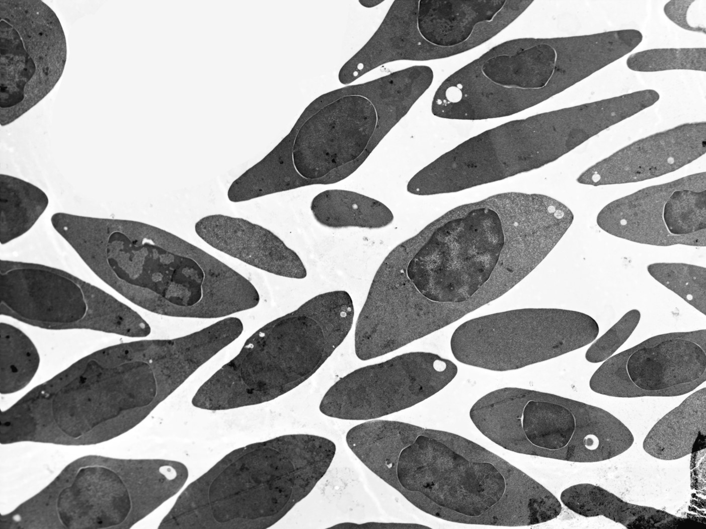
Imaging biological materials can be challenging. One of the biggest challenges is segmenting these low-contrast materials that exhibit only subtle changes in greyscale levels. As such, segmentation is one of the biggest challenges faced by today’s microscopists.
The ZEN Intellesis Image Analysis Bundles enable you to enhance your segmentations of complex samples with machine learning-driven segmentation. Machine learning-supported image segmentation utilizes deep learning techniques to overcome the limitations and challenges of threshold-based segmentation techniques to improve analysis productivity, reliability and repeatability.
The advantages of the ZEN Intellesis Image Analysis Bundle to enhance your segmentation of complex samples
Machine-Learning powered image segmentation to improve your segmentation models
- More powerful than classical threshold-based segmentation by combining various filters (e.g. intensity, edge detection, texture,…).
Simple and easy-to-use user interface for improved time-to-results
- Straightforward, easy-to-use workflow enables every microscope user to perform advanced segmentation tasks rapidly.
Segment data from any imaging source
ZEN Intellesis can be used on image files from any imaging solution provider to provide a seamless segmentation workflow.
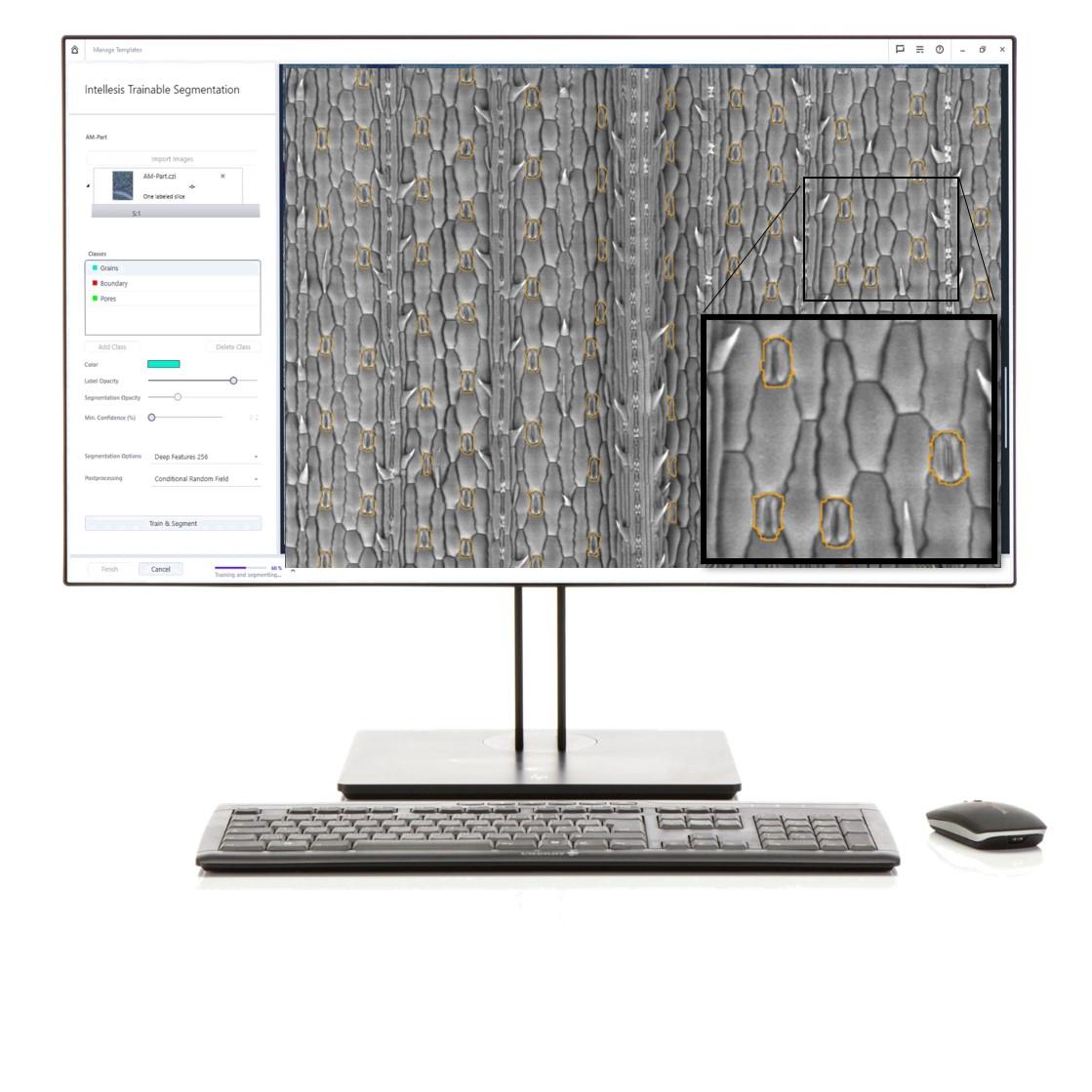

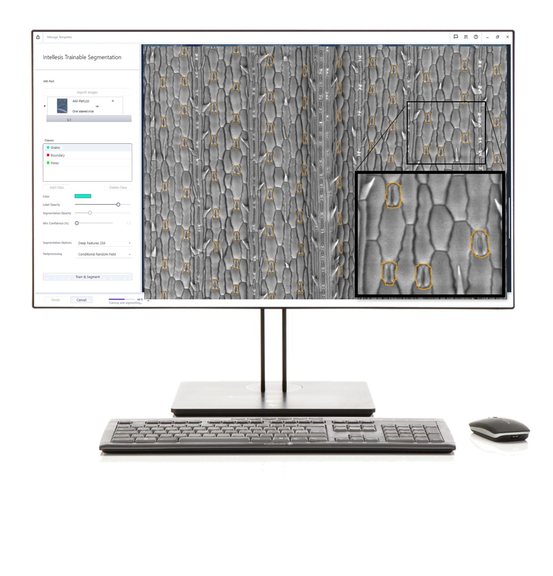

Effortlessly label and train the different objects in the ZEN Intellesis interface.
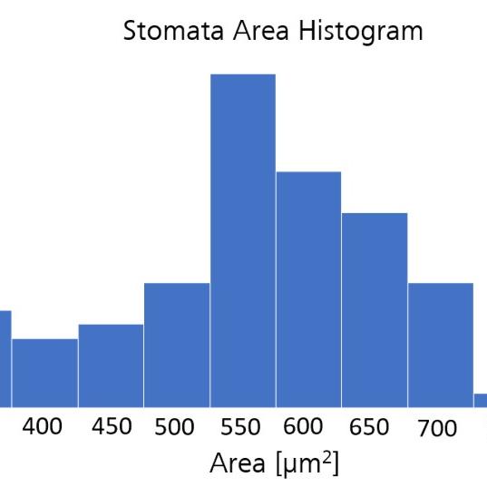


Automatic generation of statistical analysis.
Start your connected microscopy with a stereo microscope
When acquiring images of biological materials on multiple microscopes, it can be challenging to combine images and provide contextual and correlative microscopy workflows. The combination of multiple datasets provides a multi-scale and multi-model advantage to gain unique insights into your samples.
Correlative Microscopy Solution Bundle provides two imaging solutions in one correlative package. Benefit from the contextual overview images the Stemi 305 stereo microscope can provide and be used to Correlate and guide SEM image acquisition.
The advantages of the Correlative Microscopy Bundle are in multi-scale and multi-modal characterization workflows of biological samples, starting with a large overview stereo microscope image.
Easy navigation through the acquisition of low-magnification overview images
- Image your sample with ZEISS Stemi 305 stereo microscope, capture large fields of view and quickly navigate to your regions of interest (ROIs).
- Understand your sample through images in context as you zoom in and out of datasets in the correlative workspace.
- Optimal illumination through reflected light contrasts and transmitted light & capture images with ZEISS Axiocam 105 color.
Overlay and align all your images to produce a contextual sample understanding
- Load or import any image data to provide contextual sample understanding.
- Benefit from connecting complex multi-scale and multi-model data with simple overview data.
Manage your imaging project with Smart Data Management databases.
- All data is saved in a project-based database for intuitive analysis.
- Search metadata to find images and their connected database.
Easy navigation
in three steps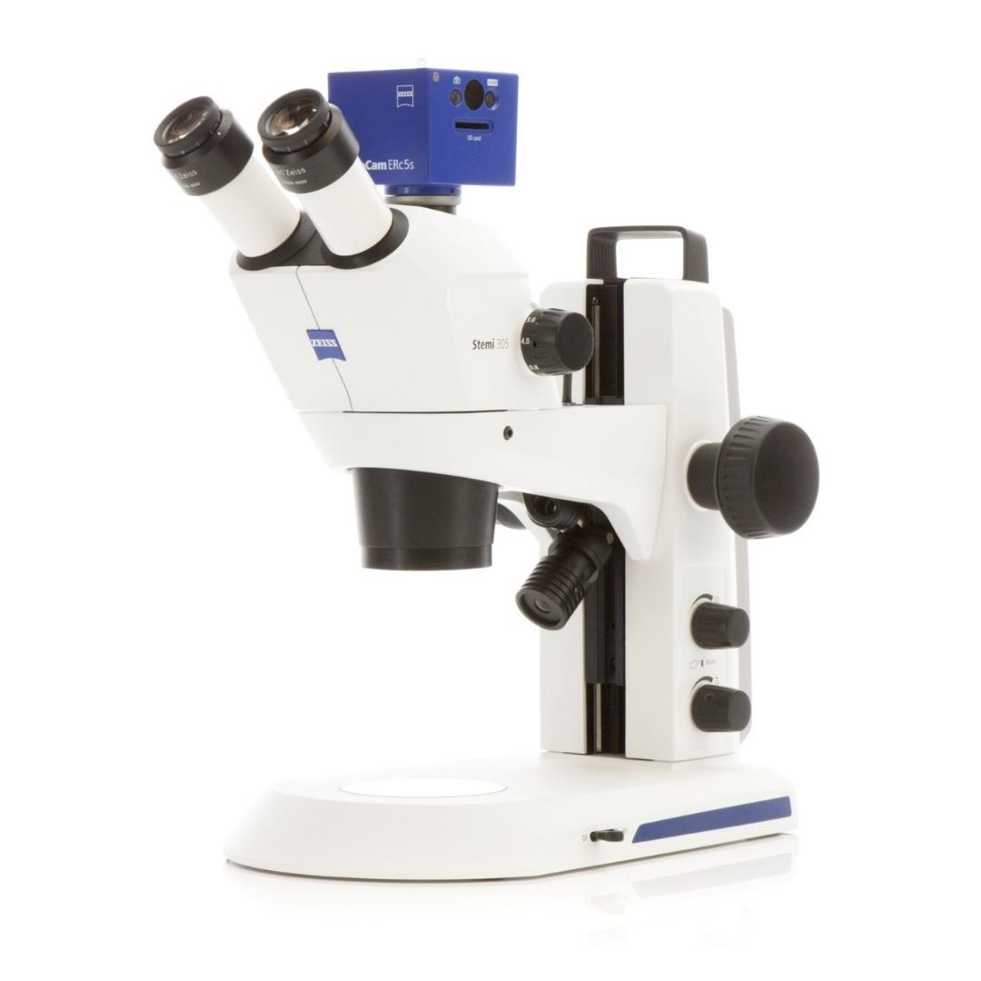


Step 1
Acquire large fields of view images using a stereo microscope.


Step 2
Acquire high-resolution images using SEM.
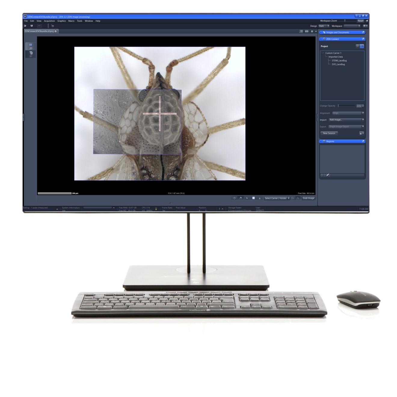

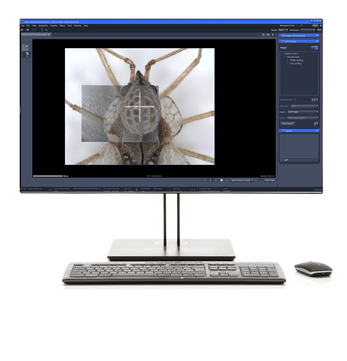

Step 3
Use the overview image to navigate and observe your high-resolution data in context.
An easy way to protect and image your hydrated samples
Hydrated and non-conductive samples provide challenges for imaging. Electron beam-induced charge artefacts impact the ability to image and understand your sample. While the vacuum conditions dehydrate samples and destroy the microstructures, you aim to image.
By combining the variable pressure mode to suppress charging artefacts and the cool stage to protect against dehydration, we provide a simple solution for biological materials imaging. The variable pressure and cool stage allow high flexibility on imaging conditions to minimize charging and simplify the complex sample preparation process.
The advantages of the Coolstage Solution Bundle in suppressing charge and protecting hydrated samples.
Overcome charging challenges
- Suppress charging artefacts for non-conductive biological samples under low vacuum conditions.
Maintain samples in a hydrated state
- Image and maintain your hydrated samples just above 0 degrees Celcius with the temperature control functions of the cool stage.
Simple sample preparation
- Save time and benefit from the simple sample preparation process without needing chemical fixation, dehydration and conductive coating.


Easily set up the cool stage to start imaging.
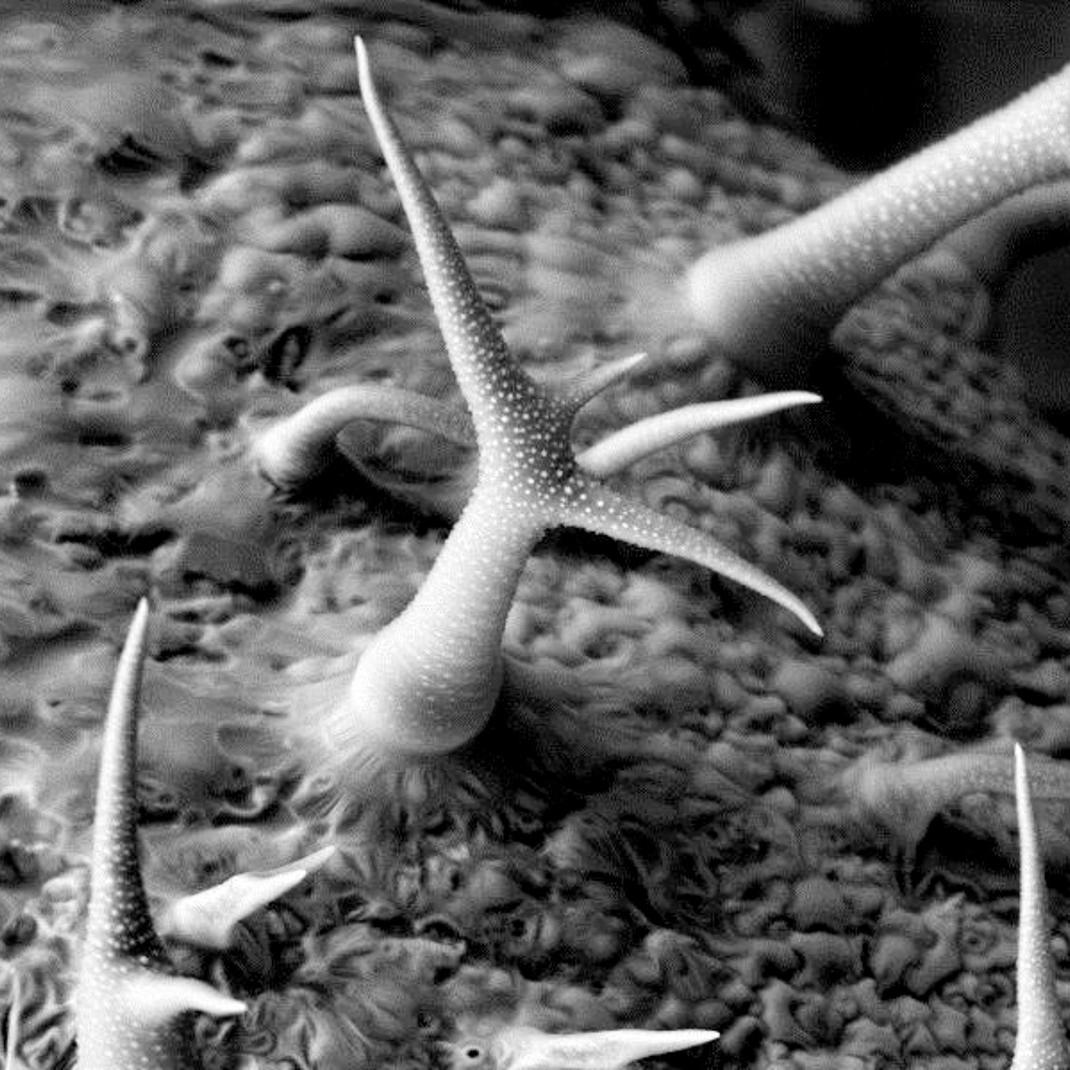

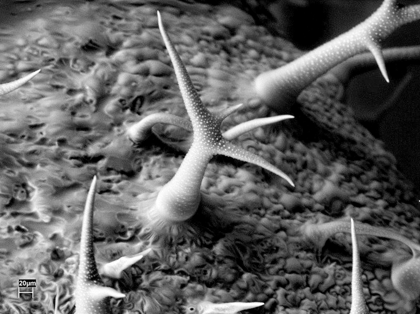

VP imaging of Trichromes on a rosemary leaf, VPSE, 20 kV, 10 Pa.
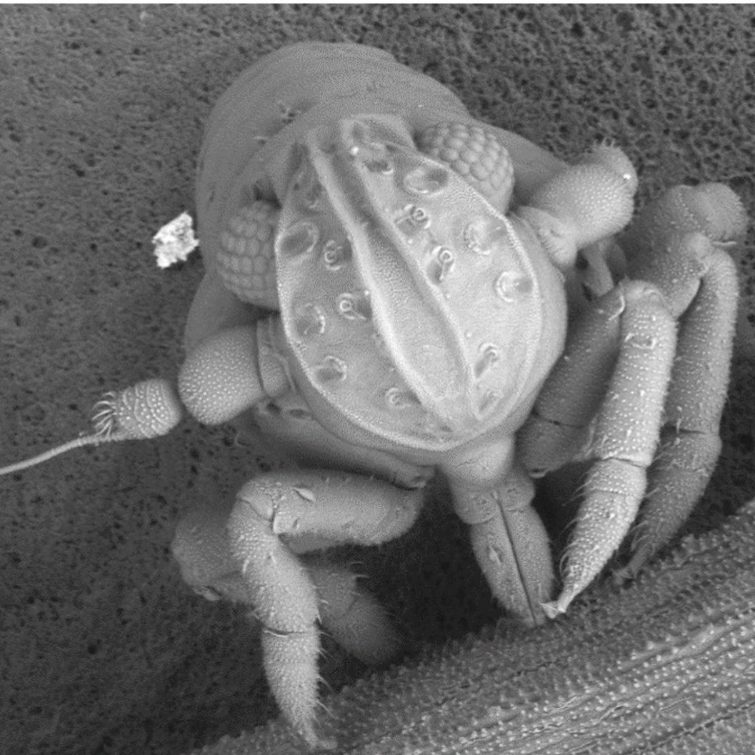

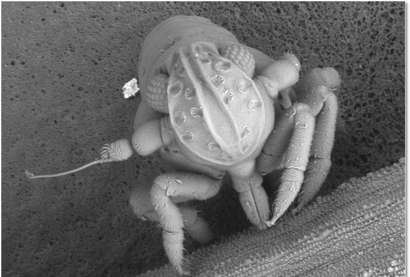

VP imaging of a louse, HDBSE, 12 kV, 60 Pa.
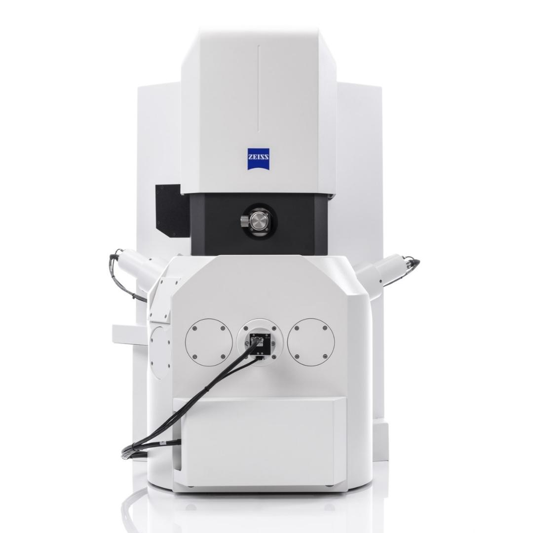
Scanning Transmission Electron Microscopy for cellular ultrastructures
Cellular ultrastructures provide clear indicators for the processes that occur in cells. While typically carried out in high-performing Transmission Electron Microscopes (TEMs), Scanning Transmission Electron Microscopy (STEM) in the SEM can provide a cost-effective, easy-to-use and quick investigation step to screen samples before TEM imaging.
Acquire cellular ultrastructure images with the EVO STEM accessory and benefit from increased imaging quality and resolution
Quickly and easily screen thin sections before TEM analysis
- Save valuable time in acquiring ultrastructure images in transmission mode.
- Perform quick screening on thin sections before TEM imaging.
Benefit from improved contrast in imaging cellular ultrastructure
- Acquire high-quality images and improved contrast in STEM mode thanks to significantly higher electron beam energies compared to conventional SEM operation.
Accessible option for transmission electron imaging
- Affordable screening tool for advanced electron microscopy workflows.


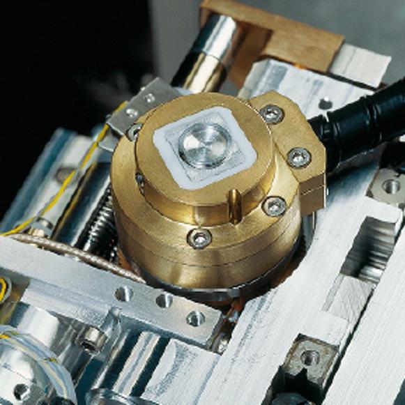
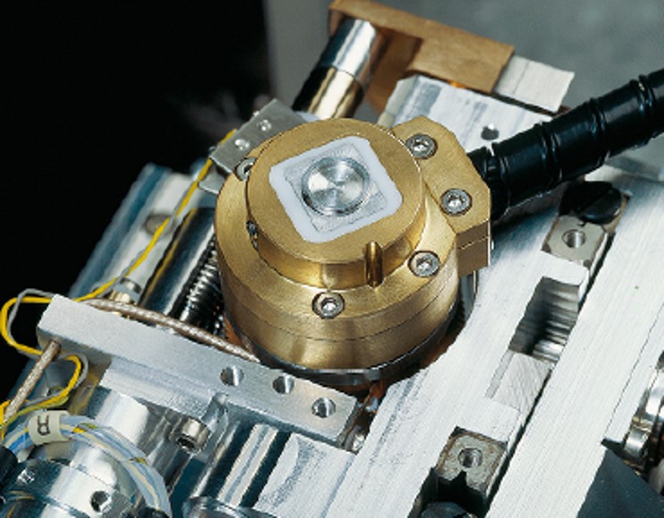
Sample placement with respect to STEM detector.
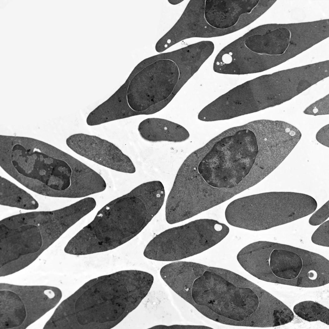


Acquire high contrast images of Python blood cells with STEM detector.
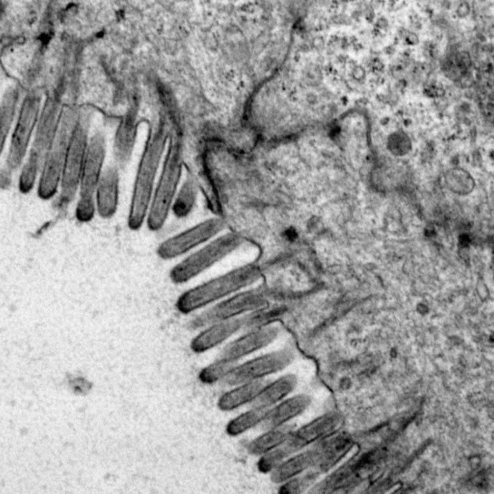

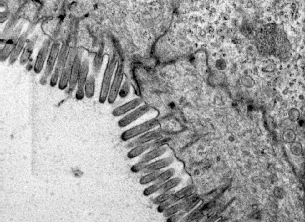

Easy imaging of sectioned intestine with microvilli border.
Keep your pharmacology workflows traceable with the GxP software
Concern over digital data integrity is everywhere, with microscopy being no exception. The GxP module in ZEN core meets the requirements of regulated industries, such as pharmaceutical or food, and helps you ensure your systems comply with FDA CFR 21 Part 11 requirements. So, when you select the EVO, you choose the microscope that is already prepared for a more regulated future.
The GxP bundle enables traceable workflows by seamlessly integrating microscopy hardware and software to meet regulated industry requirements
Keep a traceable Audit Trail
- Reproducibility and traceability of end-result relevant actions. The Audit Trail is checksum protected and prohibited from change.
Meet regulated industry standards and compliance in Pharmacology
- Pharmaceutical companies can benefit from GxP, which supports FDA 21 CFR Part 11 compliance when combined with the required qualification and validation activities.
User Management
- Configure user privileges as part of the user role management capability and ensure only users with the relevant skills can use certain functionalities.
Release Procedure Workflows
- Workflows can be set up with predetermined parameters to optimize repeatable workflows. In addition, checksum protection of data and electronic signatures ensures no changes can be made in released product workflows.
Disaster Recovery
- Backup and restore functionality ensures a short-time production-ready state in case of system breakdown.


The GxP Module offers all functionalities needed for FDA 21 CFR Part 11 compliance, such as electronic signatures.
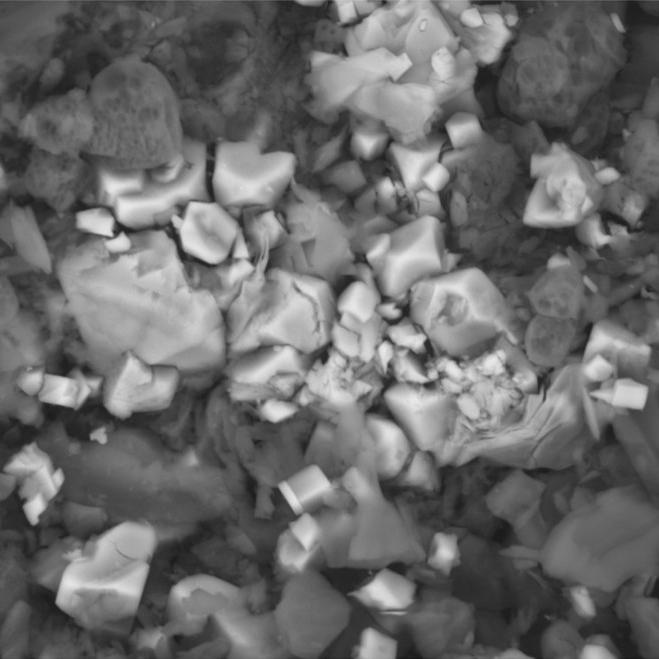

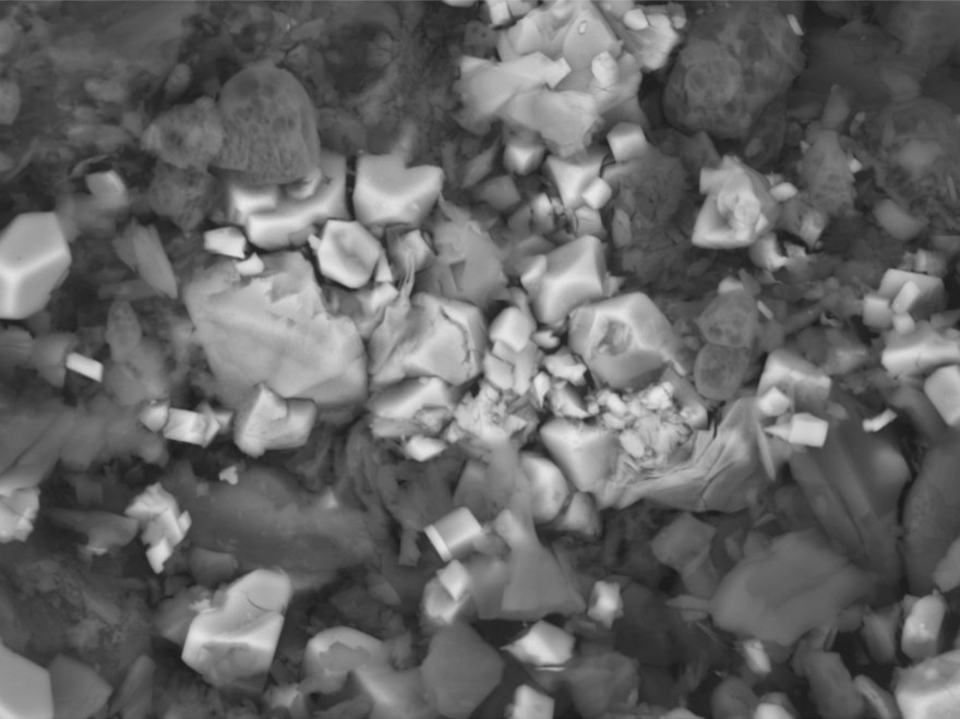

Imaging of Aspirin drug particle in GxP workflow.
Sign up to view an on-demand webinar!
Connect with a ZEISS ExpertIn this webinar, we will introduce the solution bundles specifically for life sciences applications, which greatly benefit researchers in routine workflows and gain unique insights into biological samples to answer scientific questions.