ZEISS EVO Solutions for New Materials

ZEISS EVO Solutions for Electronics
The growing trends in 5G, IoT, and AI drive consumer demand for high performance in electronic devices and components. As a result, manufacturers strive to achieve higher yields and defect-free chips. Therefore, scanning electron microscopy for visual inspection and composition analysis of surface features and defects in integrated circuits, semiconductor devices, LED, sensors, and electronic packages has become routine and essential.
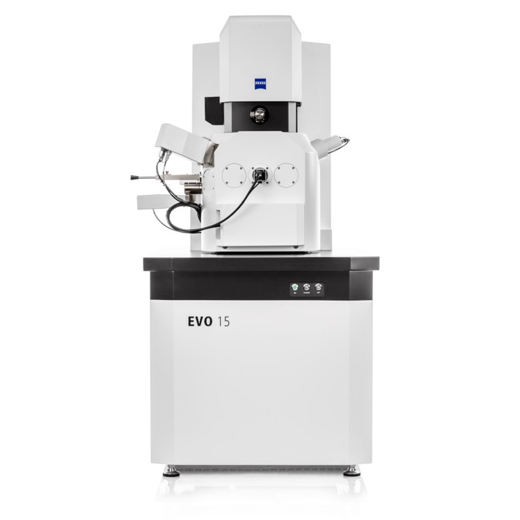
The EVO series is a high-performance scanning electron microscope with an intuitive, user-friendly interface to provide fast, reliable results. The preconfigured microscopy systems are flexible, with upgrade-friendly options and modular features for various applications. This includes automated workflows to deliver high productivity and fast time to result with simplified operation, drastically reducing training costs.
The ZEN software suite combines images from different modalities for user-customized analysis, leveraging advanced image processing and data analysis functions to provide insights and actionable information. In addition, smart and automated features enable the automation of repetitive analysis tasks and reduce complex image segmentation and measurements of features in a simple, user-friendly manner, improving productivity and time to results at an affordable cost.
With the ZEISS ZEN software suite, EVO "solution bundles" provide electronics and semiconductor solutions to address routine inspection, advanced automated materials/device analysis, critical features, component measurement and metrology.
The tabletop SEM alternative for high-performance imaging and data analysis
To characterize New Materials, producing images is only part of the workflow. The imaging of microscope features is critical in understand a materials fundamental properties. However to characterize these new materials, imaging is only part of the workflow.
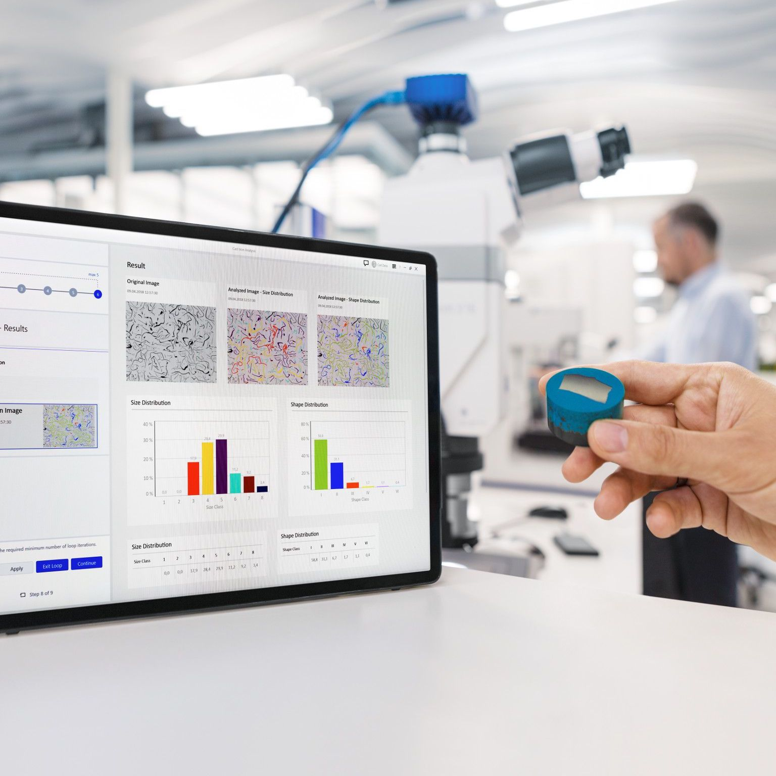
The ZEN Imager Analysis bundle allows you to generate automatic measurements. Pre-defined measurement parameters and workflows enable fast and simple analysis using the online analysis mode.
The advantages of the ZEN Image Analysis bundle to improve automation and repeatability of image segmentation and analysis workflows
Benefit from automated and repeatable workflows
- Using the measurement program wizard, you can tackle complex measurement tasks in just a few minutes.
- Once created, the programs are always available, and you can use them to analyze unlimited images.
Take advantage of the full flexibility to define and control the measurement process
- Retain full control over the measurement process and adjust the settings to your needs.
Automated report generation for improved productivity
- Use ZEN core´s powerful reporting functionality to create reports in MS Word or PDF format.
- Fully automate the reporting process with ZEN core’s Job Mode.
Application in Focus – Additive Manufacturing
Easy navigation
in four stepsAutomatic segmentation of the brighter phase corresponding to particles.
Automatic generation of reports and statistical analysis.
Using the power of machine learning for image segmentation and analysis
Image segmentation is the foundation of all image analysis steps. Image segmentation is one of the biggest challenges in microscopy when characterizing new materials. Compromising your image segmentation also means compromising the reliability of the analysis and subsequent results.
The ZEN Intellesis Image Analysis bundle enables you to enhance your segmentations of complex samples with machine learning-driven segmentation. Machine learning-supported image segmentation utilizes deep learning techniques to overcome the limitations and challenges of threshold-based segmentation techniques to improve analysis productivity, reliability and repeatability.
The advantages of the ZEN Intellesis Image Analysis bundle to enhance your segmentation of complex samples
Machine Learning image segmentation module
- More powerful than a classical threshold-based segmentation through combining a variety of different filters (e.g. intensity, edge detection, texture)
Simple and easy-to-use user interface for improved time-to-results
- Straightforward, easy-to-use workflow that enables every microscope user to perform advanced segmentation tasks rapidly
Segment data from any imaging source
- ZEN Intellesis can be used on image files from any imaging solution provider to provide a seamless segmentation workflow
Application in Focus – Ceramics
Effortlessly label and train the different objects in the ZEN Intellesis interface.
Full integration in the image analysis module allows job mode and automation tools.
The same image is segmented using traditional threshold methods giving poor segmentation due to similar greyscale values for different constituents.
Connect multiple perspectives of your samples across different imaging techniques and modalities
You must combine multiple microscopy technologies or SEM modalities to understand your New Materials samples fully. This often includes time-consuming workflows to locate regions of interest for further imaging and analysis.
The ZEN Connect Solution bundle allows you to connect multiple perspectives of your sample across scales and imaging modalities. In addition, the ZEN connect Solution bundle enables you to bring all imaging technologies together – ZEISS or not – to answer your research questions.
The advantages of ZEN Connect in multi-scale and multi-modal characterization workflows of New Materials samples
Easy navigation through the acquisition of low-magnification overview images
- Image your sample with any low magnification system and use it to navigate to your regions of interest (ROIs).
- Understand your sample through images in context as you zoom in and out of datasets in the correlative workspace
Overlay and align all your images to produce a contextual sample understanding
- Load or import any image data:
- Benefit from connecting complex multi-scale and multi-model data with simple overview data
Manage your imaging project with Smart Data Management databases
- All data is saved in a project-based database for intuitive analysis.
- Search metadata to find images and their connected database
Application area in Focus - Magnetic Materials
Easy navigation
in four stepsStep 1
Use your favourite low-magnification system to acquire large fields of view.
Step 2
ZEN Connect organizes your images in a well-defined project.
Step 3
Align your high-resolution system to the overview image.
Step 4
Use the overview image to navigate and observe your high-resolution data in context.
Increasing productivity and ease of use in your light-to-electron microscopy workflow
Once you have identified one region or location of interest using light microscopy, the natural workflow is to move the samples to the SEM to perform higher-resolution imaging or chemical analysis. It remains challenging to relocate this specific region of interest once the sample has been transferred from the light microscope to SEM.
The ZEN Connect Correlation Solution Bundle allows you to locate these regions of interest directly. This correlative workflow enables the correlation of stage coordinates to provide a fast and efficient workflow to locate features between instruments.
The advantages of the ZEN Connect Correlation Solution Bundle in improving workflow productivity and ease of use between multiple microscopes
Fast calibration for easy navigation
- Instead of wasting valuable time relocating regions of interest from microscope-to- microscope, you can now calibrate and navigate swiftly with only a few mouse clicks
Correlative samples holder for precise relocations
- Semi-automatic 3-point calibration of reference markers allows for precise relocation to regions or features of interest
- Create correlative data overlays to improve sample understanding and reporting
Application area in Focus - Additive Manufacturing


Use your favourite low-magnification system to acquire large fields of view.

ZEN Connect organizes your images in a well-defined project.
Automatic relocation to precipitate, followed by high-resolution image acquisition and overlay over a large field of view light microscope image.
Start your Connected Microscopy processes with a stereomicroscope and EVO workflow
Often to fully understand your New Materials samples, you will need to combine light and electron microscopy images. This usually includes time-consuming workflows to locate regions of interest for further imaging and analysis.
Correlative Microscopy Solution Bundle allows you to capture a large field of view stereomicroscope image of your sample under various illumination methods and connect it to the high-resolution SEM images for easier navigation and contextual understanding.
The advantage of the ZEN Correlative Microscopy bundle is in multi-scale and multi-modal characterization workflows of New Materials samples, starting with a large overview stereomicroscope image.
Easy navigation through the acquisition of low-magnification overview images
- Image your sample with ZEISS Stemi 305 stereo microscope, capture large fields of view and quickly navigate to your regions of interest (ROIs).
- Understand your sample through images in context as you zoom in and out of datasets in the correlative workspace
- Optimal illumination through reflected light contrasts and transmitted light & capture images with ZEISS Axiocam 105 color
Overlay and Align all your images to produce a contextual sample understanding
- Load or import any image data to provide contextual sample understanding
- Benefit from connecting complex multi-scale and multi-model data with simple overview data
Manage your imaging project with Smart Data Management databases
- All data is saved in a project-based database for intuitive analysis.
- Search metadata to find images and their connected database
Application area in Focus - Magnetic Materials
Easy navigation with scientific context
Easy navigation
in three stepsStep 1
Acquire large fields of view images using the stereo microscope.

Step 2
Acquire high-resolution images using SEM.
Step 3
Use the overview image to navigate and observe your high-resolution data in context.
Sign up to view an on-demand webinar!
Connect with a ZEISS ExpertIn this webinar, we will outline the power of the ZEN ecosystem showing how its multi-scale and multi-modal analysis capabilities are used in the characterization of new materials.