

Revolutionize Palm Oil Research and Quality Control with ZEISS Sigma
Empowering palm oil professionals with cutting-edge imaging technology to optimize oil yield, ensure quality, and accelerate innovation.
Why ZEISS Sigma with Gemini Technology for Palm Oil Applications?
Short description of the module

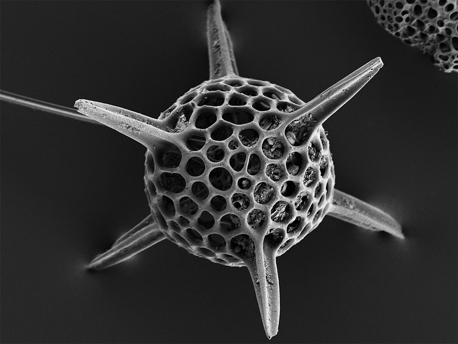
Unmatched Cellular Imaging for Breeding Innovation
Visualize and analyze palm fruit cellular structures in unparalleled resolution to identify high-yield variants. ZEISS Sigma's Gemini optics deliver precise imaging even at challenging working distances, empowering breeding innovation.


Elevated Quality Control
Ensure top-tier palm oil purity with ZEISS Sigma. The Gemini optics’ optimized detection efficiency captures precise microstructural details, allowing you to identify and eliminate impurities with confidence.


Optimize Soil and Fertilizer Management
Maximize plantation efficiency by understanding soil microstructures with ZEISS Sigma. The intelligent imaging capabilities of Gemini optics provide the insights you need to make data-driven decisions for sustainable nutrient management.


Accessories


In Situ Lab for ZEISS FE-SEMs
Link Materials Performance to MicrostructureExtend your ZEISS FE-SEM with an in situ solution for heating and tensile experiments. Benefit from an integrated solution. Investigate materials like metals, alloys, polymers, plastics, composites, and ceramics. Combine a mechanical tensile or compression stage, a heating unit and dedicated high-temperature detectors with analytics. Control all system components from a single PC with a unified software environment that enables unattended automated materials testing.


SmartEDX
Discover Embedded Energy Dispersive X-ray Spectroscopy

Fully Integrated RISE
Benefit from Raman Imaging and Scanning Electron MicroscopyComplement the characterization of your material and add Raman Spectroscopic Imaging (RISE). Get a chemical fingerprint from your sample and extend your Sigma 300 with confocal Raman imaging capability. Recognize molecular and crystallographic information. Perform 3D analysis and correlate SEM imaging, with Raman mapping and EDS data if appropriate. Fully integrated RISE lets you take advantage of both best-in-class SEM and Raman systems.
Ready to Take Palm Oil Innovation to the Next Level?
Request a Consultation TodayLet’s discuss how ZEISS Sigma can empower your palm oil research and production.