Register For ZEISS's X-ray Microscopy Webinar Series Below
Learn How ZEISS X-Ray Microscopy Technologies Can Help Your Materials Science Research

Sign up for free to view the webinars!
Join our free webinar series below with Microscopy expertsLaboratory-based X-ray Microscope (XRM) instruments are becoming a “must-have” for advanced non-destructive multi-scale material characterization in 3D and 4D. The ZEISS Xradia Versa portfolio provides a versatile, industry best-resolution and contrast platform to allow you to expand the boundaries of your non-destructive sub-micro scale imaging.
This webinar series will summarize the ZEISS Xradia Versa XRM technology, its capabilities, and how the XRM can be used in a range of different applications and workflows. This webinar will include a range of workflows involving the ZEISS LaserFIB to access deeply buried structures, 4D in-situ experiments, and Diffraction Contrast Tomography for crystallographic imaging. The series will explore application examples in the area such as battery materials, electronics, ceramics, additive manufacturing, metals, and alloys.
An Introduction to 3D and 4D Imaging with the ZEISS Xradia Versa Bringing Synchrotron Capabilities to the Lab with X-ray Microscopy
Breakthrough innovations in source and optics technology have extended the limits of what is possible with laboratory-based 3D and 4D non-destructive x-ray imaging. The ZEISS Xradia Versa is a “X-ray Microscope” (XRM) that provides a class above traditional microCT systems. Through a combination of a unique two-stage magnification optics and a high flux higher source, the ZEISS Xradia Versa is able to produce faster sub-micron scale resolution imaging across the widest range of sample sizes.
In this webinar, we will focus on introducing the technology and the benefits it provides using a wide range of application examples in Material Science, Life Science and Electronics fields.
Key Learnings
- An understanding of the concept of X-ray Microscopy (XRM)
- An understanding of the advantages XRM provides in 3D non-destructive X-ray imaging in samples size, resolution, contrasts and throughput
- An understanding of some of the applications that XRM can address (i.e., in-situ testing, composites, batteries, additive manufacturing etc.)
How X-ray Microscopy and “Resolution at a Distance” (RaaD) can overcome the sample size vs resolution limitations of microCT
Non-destructive 3D techniques have provided a leap forward in capabilities available to researchers. These microCT or X-ray Microscopy techniques allow researchers to characterizes 3D objects and their internal structures. However, conventional microCT techniques have limitations when attempting to image “multi-scale” samples. Multi-scale samples typically exhibit important characteristics across lengths scales (i.e., macro, micro, nanometer).
In this webinar, we will define the “multi-scale” problem and how limitations of microCT mean resolution typically is reduced when sample size increases. We will also outline the advantages of the unique Resolution at a Distance (RaaD) capabilities of the ZEISS Xradia Versa.
RaaD capabilities enable “interior tomography” whereby high resolutions scans can be acquired from deep inside large 3D samples and overcoming the multi-scape problem associated with microCT.
Key Learnings
- An understanding of the multi-scale problem in Material Science and the challenges this bring to non-destructive 3D imaging
- An understanding of the Resolution at a Distance concept and how it overcomes the multi-scale (sample size vs. resolution) problem
- An understanding of some of the many Material Science applications that the RaaD capability in the ZEISS Xradia Versa can address
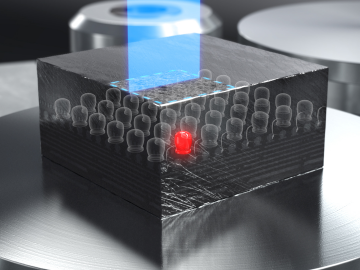
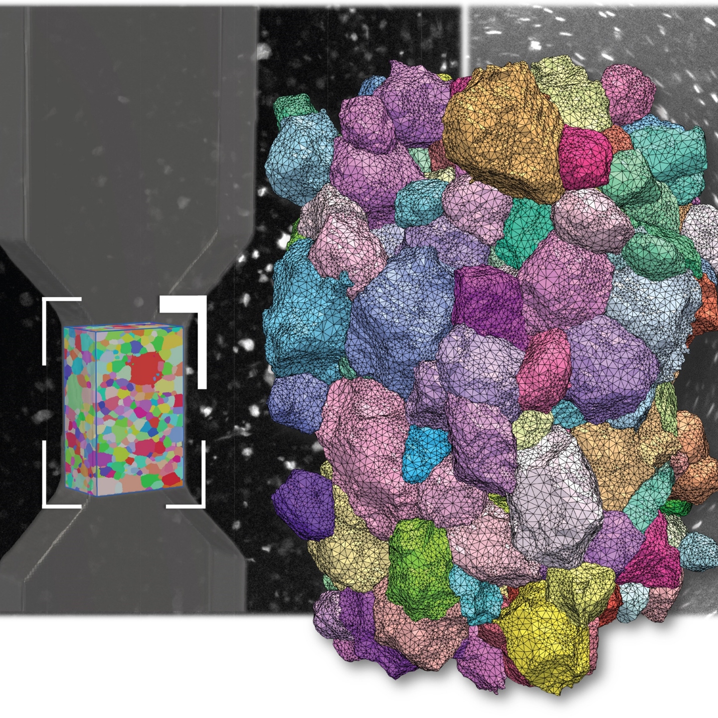
Rendered animation showing how LabDCT works
Achieve direct visualization of 3D crystallographic grain orientationAdvances in X-ray Microscopy on Crystallographic Imaging of Polycrystalline Materials Lab-based diffraction contrast tomography
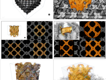
X-ray tomography for 3D non-destructive imaging has been widely adopted and operated under two primary contrast mechanisms: X-ray absorption and phase contrast. Both these contrast mechanisms rely on material density differences. However, when imaging polycrystalline materials (i.e., metals and alloys, ceramics etc.) these contrast mechanisms become ineffective. For two decades synchrotron-based Diffraction Contrast Tomography (DCT) methods have been able to overcome this challenge and provide “crystallographic contrast” by using the diffraction signals of polycrystalline samples.
In this webinar we will outline Diffraction Contrast Tomography (DCT) method and how it is available in a laboratory-based DCT system. We will explore the latest advances in diffraction scanning modes that address challenges of achieving true sample representivity by investigating massive volumes of samples at an unprecedented scale. We will further demonstrate applications use cases of extremely large volume grain mapping on a wide variety of materials ranging from metals, ceramics and geomaterials. Finally, we will present some unique correlative workflows spanning across X-ray microscopy, non-destructive grain mapping combined with high resolution scanning electron microscopy to get valuable insights into the multiscale material microstructure.
Key Learnings
- Understand the Diffraction Contrast Tomography (DCT) techniques can be used on a laboratory-based system
- Understand the significance of true sample representatives and how new non-destructive methods and cadres this critical challenge
- How the destructive and non-destructive approaches can be combined to gain unique insights into a materials science sample
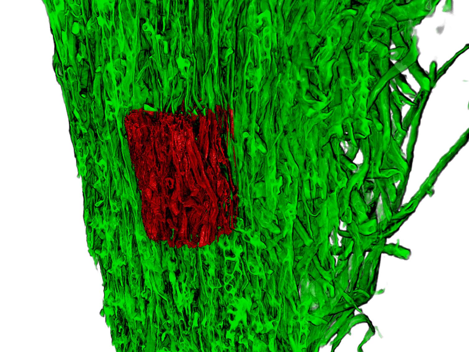
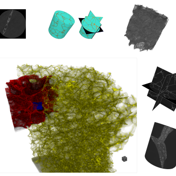
Correlative study to examine corrosion damage in a magnesium alloy
Video courtesy of Philip Withers, Henry Moseley X-ray Imaging Facility , The University of Manchester.Correlative Microscopy move beyond the boundaries of microscopy to achieve the best in class results
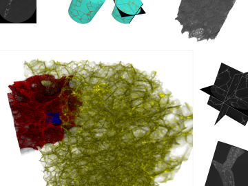
To capture the essence of your sample, you often need to analyze it with more than one method. Going from the micro to nano scale can require correlating light with electron microscopy (CLEM), or X-ray with FIB-SEM (CXF). Correlative microscopy from ZEISS gives you integrated solutions and seamless workflows. Decide on ZEISS as your sole provider of light, electron, ion and X-Ray microscopes and profit from the long years of experience in correlative analysis. Choose unique sample centric correlation of images and data to advance your work beyond the limiting boundaries of a single microscopy technique.
Key Learnings
- An understanding of the multi-scale challenges we face in Material Science and the need for workflows that can bridge across multiple-length scales (mm to nm)
- An understanding of the ZEISS X-ray Microscope (XRM) to LaserFIB workflow in identifying and accessing deeply buried microstructures
- An understanding of a number of applications that this workflow can be applied to (i.e., batteries, electronics, metals etc.)
Extending the Throughput and Performance Limitation for Sub-Micron Imaging of Intact Samples
ZEISS Xradia Versa 3D X-ray microscopes (XRM) use the combination of resolution and contrast with flexible working distances to extend the power of non-destructive imaging in your lab; Benefit from its architecture that uses a two-stage magnification technique to achieve submicron resolution at a distance (RaaD). Reducing dependence upon geometric magnification maintains submicron resolution even at large working distances.
In this webinar, we will unlock new degrees of versatility for your scientific research with the ZEISS Xradia Versa 3D X-ray microscopes (XRM). Discover how the speed of data acquisition and reconstruction, as well as image quality, are enhanced without sacrificing resolution by both hardware and software. Boost the performance of the Xradia Versa by exploring advanced capabilities. Discern your materials with Xradia synchrotron-caliber optics to maximize the throughput and image quality performance.
Key Learnings
- Understand the variety of opinions for improving the speed of the data acquisition and image quality
- New reconstruction technologies enabling throughput or image quality improvements in 3D X-ray imaging
In Situ & 4D Science Observing and Quantifying the Evolution of 3D Microstructure
Since the advent of the microscope, engineers and scientists have refined imaging techniques to observe 2D and 3D microstructures and quantify their evolution over time, or as the result of external stimuli such as temperature, force, etc. The vast majority of 3D characterization techniques require samples to be destructively cut or consumed, rendering observation under realistic conditions over time impossible. ZEISS 3D X-ray microscopes (XRM) are uniquely suited to image materials in situ under variable environments in 4D (3D plus time) in order to non-destructively characterize and quantify the evolution of 3D microstructures. From materials testing under mechanical load or at elevated temperatures, to observing flow in porous media, ZEISS environmental sample holders and innovative features such as “resolution at a distance” (RaaD) facilitate a broad range of in situ and 4D research and discovery.
Key Learnings
- Understand the unique strength of RaaD techniques for non-destructive characterization of microstructures in controlled environments and overtime


