Introducing ZEISS Sample-in-Volume Analysis workflow
Identify, Access, Prepare, Analyze your sample with precise navigational guidance
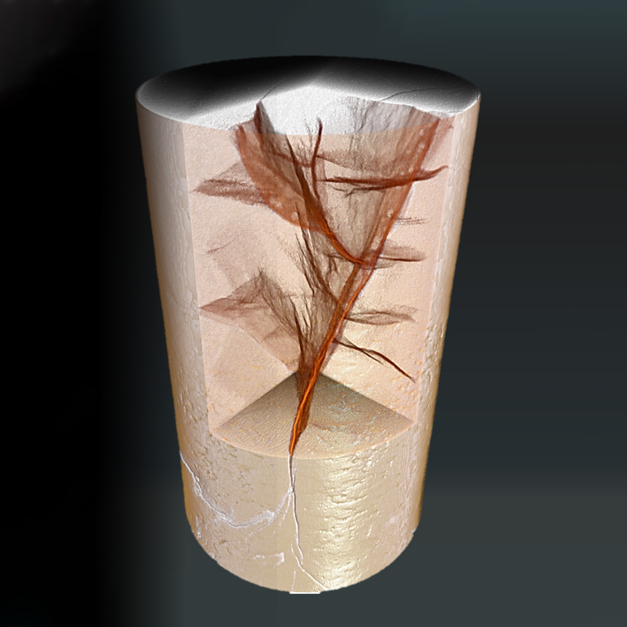
Understanding the multi-scale microscopy challenge
For Material Scientists
Do You Struggle To...
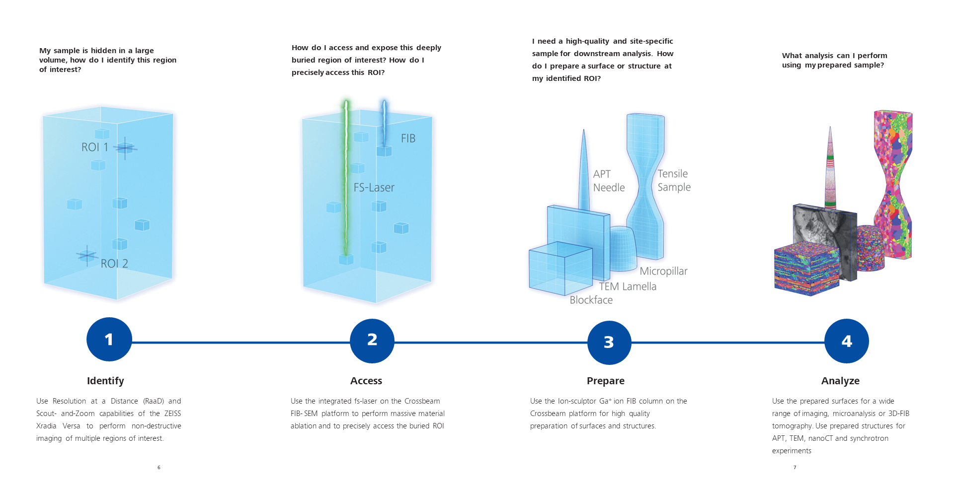
• Identify deeply buried defects or samples in large volume?
• Precisely access Regions-of-Interest (ROI) within a volume for sample preparation such as TEM, APT or microanalysis samples?
• Establish multi-scale or multi-dimensional information ranging from macro- to sub-nanometer length scale?
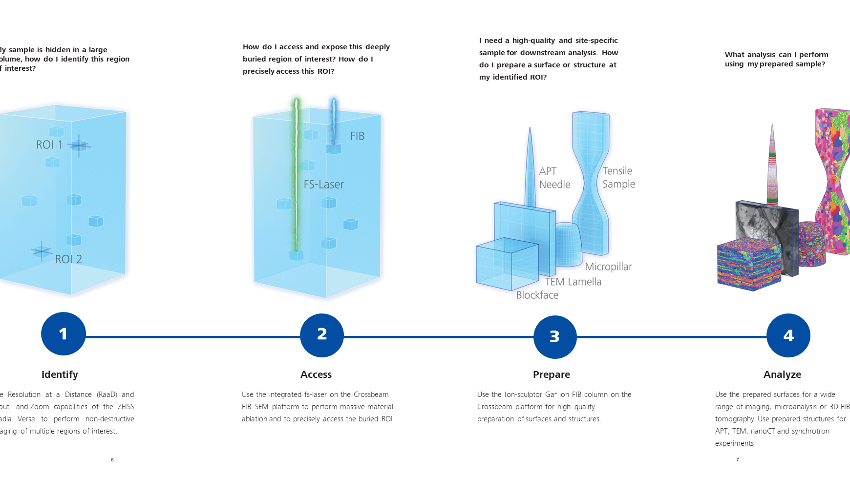
Here's What You'll Learn In The SamVol Webinar
Characterization of samples in material science requires an understanding of structures, processes and properties across different length scales. As we move from the macro-scale towards sub-nanometer, we need a workflow that enables us to make the best decisions possible for the best experimental outcomes by:
- Helping to identify regions of interest in large samples
- Provide a method to precisely access regions buried within our samples that contain the nm-scaled information required
- A preparation step that allows us to create high-quality surfaces and structures for further analysis
ZEISS has developed a unique correlative workflow to address these multiscale challenges. This workflow is known as the Sample-in-Volume Analysis workflow and synergizes the latest innovations in multi-scale microscopy.
Who is this webinar for?
If you are a material scientist, research engineer, microscopist or technologists who wants to learn about ZEISS multi-scale microscopy solutions, this webinar is for you.
Here's what you'll learn in the free webinar:
• The ZEISS multi-scale microscopy workflow that characterizes length scales from macro- to sub-nanometer
• How 3D non-destructive X-ray microscopy can help identify regions of interest for downstream experiments
• How the ZEISS Crossbeam platform with the integrated fs-laser provides a paradigm change for ion beam microcopy with the capability to achieve massive material ablation to access deeply buried structures
• How the Ion-sculptor provides high quality surfaces and structures for further experiments and analysis
• How the ZEISS Atlas 5 software can provide the correlative link to allow precise and site-specific access to regions of interest
Video of the Sample-in-Volume Analysis workflow
Your Sample-in-Volume with the Versa 3D X-ray Microscope (XRM)
The ZEISS Xradia Versa enables non-destructive 3D X-ray microscopy (XRM) for imaging of large samples. The synchrotron-like optics provide two distinctive advantages in addressing the multi-scale microscopy challenge:
- Use Resolution at a Distance (RaaD) technology to maintain a high spatial resolution while imaging large sample volumes
- Benefit from the Scout-and-Zoom capability to locate multiple regions of interest and perform higher resolution non-destructive “interior tomography” for region of interest identification
Download the brochure to learn more about the workflow and example applications in catalysts & nanoparticles, battery, metals & additive manufacturing, geoscience and electronics and device research.
Your Sample-in-Volume with the LaserFIB for ZEISS Crossbeam
The LaserFIB for the ZEISS Crossbeam integrates a femtosecond laser onto the ZEISS Crossbeam (FIB-SEM) platform. The integrated laser enables massive and rapid material removal. This enables you to mill through large volumes and access the identified regions of interest buried deeply within your sample.
- ZEISS ATLAS 5 allows you to correlate your XRM data for precise targeting of regions of interest for LaserFIB milling
- Prepare large cross sections and trenches to access your deeply buried region of interest in minutes.
- Protect your FIB-SEM from contamination by performing the laser milling in a dedicated and isolated load lock chamber
Download the brochure to learn more about the workflow and example applications in catalysts & nanoparticles, battery, metals & additive manufacturing, geoscience and electronics and device research.
Your sample surface or structures for further analysis
The ZEISS Crossbeam with the Ion-sculptor Ga+ ion column can provide you with exceptional surfaces finishing to prepare delicate structures and high-quality surfaces.
- Perform live imaging and milling to provide precise navigational guidance to the region of interest
- Use the Ion-sculptors low voltage capabilities for fine polishing and the preparation of ultra-thin samples
- Benefit from the 100 nA probe current to ensure fast and precise preparation without compromising on resolution
Download the brochure to learn more about the workflow and example applications in catalysts & nanoparticles, battery, metals & additive manufacturing, geoscience and electronics and device research.
Your Prepared Sample
The ZEISS Crossbeam platform allows you to configure a range of analytical modalities on to the Crossbeam platform. Take the prepared sample to external instruments (i.e., TEM, APT) to extend the Sample-in-Volume Analysis to the sub-nanometer
- Perform microanalysis using on your identified region of interest (i.e., EDX, EBSD)
- Analyze trace elements with ToF-SIMS attached to the Crossbeam platform for a completely air free workflow
- Benefit from ZEISS Atlas 5 for 3D FIB tomography, 3D EBSD and 3D EDS analysis capabilities
Download the brochure to learn more about the workflow and example applications in catalysts & nanoparticles, battery, metals & additive manufacturing, geoscience and electronics and device research.
Webinar Agenda
Time |
What We Are Going To Cover |
|
---|---|---|
40 Minutes |
|
|
20 Minutes |
Panel discussions Discuss with ZEISS scientists and technologists on the workflow and its application examples |
|
10 Minutes |
|
|
10 Minutes |
Technology in Focus – LaserFIB for ZEISS Crossbeam & ZEISS Atlas 5 Learn how the femto-second laser integrated on the ZEISS Crossbeam platform enable massive material ablation that rapidly and precisely access samples from Regions-of-Interest (ROI) that are buried deeply in large volume. Learn how the ZEISS Atlas 5 correlative software enables precise navigation and access of ROI |
|
10 Minutes |
|
|

Your presenter for this webinar
Shaun Graham
Shaun graduated from the University of Leicester in 2013 with a masters degree in Earth Science before joining ZEISS Microscopy Customer Center in Cambridge as a Electron Microscopy application specialist. In 2015, Shaun joined the Strategic Marketing team as an application development engineer before joining the ZEISS APAC team to lead business development activities for Materials Research.