

ZEISS @ AEGC 2023
Advanced microscopy solutions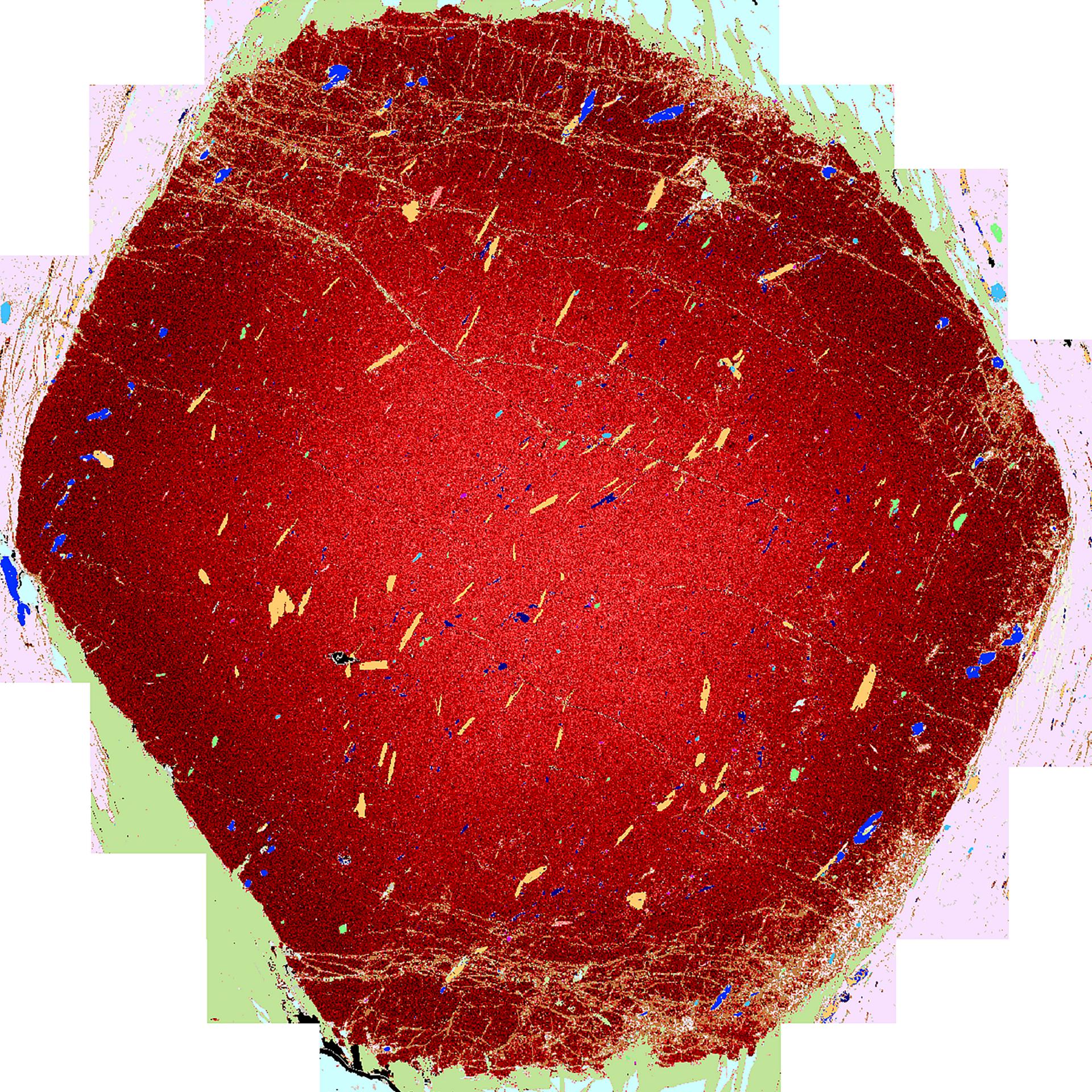

Mapping Geochemical Zoning with Quantitative EDS
ZEISS MineralogicZEISS Mineralogic (ML) is an analytical SEM with automated mineralogical mapping using quantitative geochemical analysis through Energy Dispersive Spectroscopy (EDS). ML is a EM system designed specifically to obtain quantitative chemistry and automatically classify mineral phases direct from thin sections.
The result is a unique system that can combine the two key aspects of quantitative EDS – rapid phase mapping, and geochemical analysis.
The results can be visualized as a CHEMera map, where minerals are automatically identified by ML and phases of interest have their internal geochemical zonation exposed.
ML is versatile, with a variety of imaging detectors that can be applied alongside the EDS capability. In this instance we present the capabilities of ZEISS Mineralogic based on ZEISS Sigma VP FE-SEM equipped with Back Scattered Electron (BSE) imaging and two Oxford Instruments Ultim Max 65 EDS detectors.

3D Automated Quantitative Mineralogy
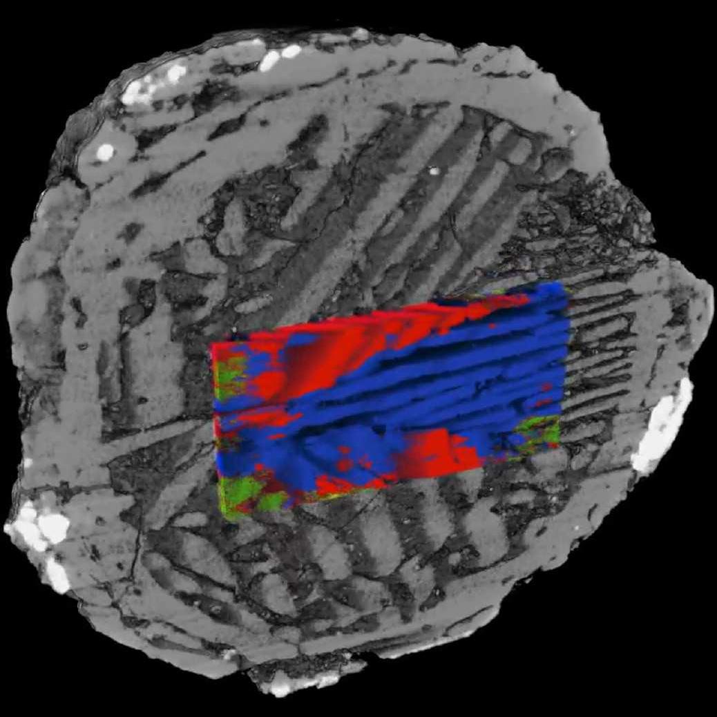
ZEISS MineralogicRock and mineral assemblages are three dimensional (3D) structures, but traditional microanalytical techniques used to examine and address them, such as scanning electron microscope (SEM) based automated quantitative mineralogy (AQM), or traditional optical petrography, are inherently 2D. The prevalence of these techniques is understandable as they give a great level of analytical precision and reliability, but their 2D nature limits the insights that are available to the researcher, as well as their ability to make quantitative assessments.
X-ray microscopy is a technique whereby 3D volumes can be reconstructed from a set of projections using back-projection. The greyscale value of a voxel (3D pixel) within these volumes is proportional to the relative X-ray attenuation within that voxel, which is in turn related to the constituent material density, atomic weight and the incident X-ray energy.