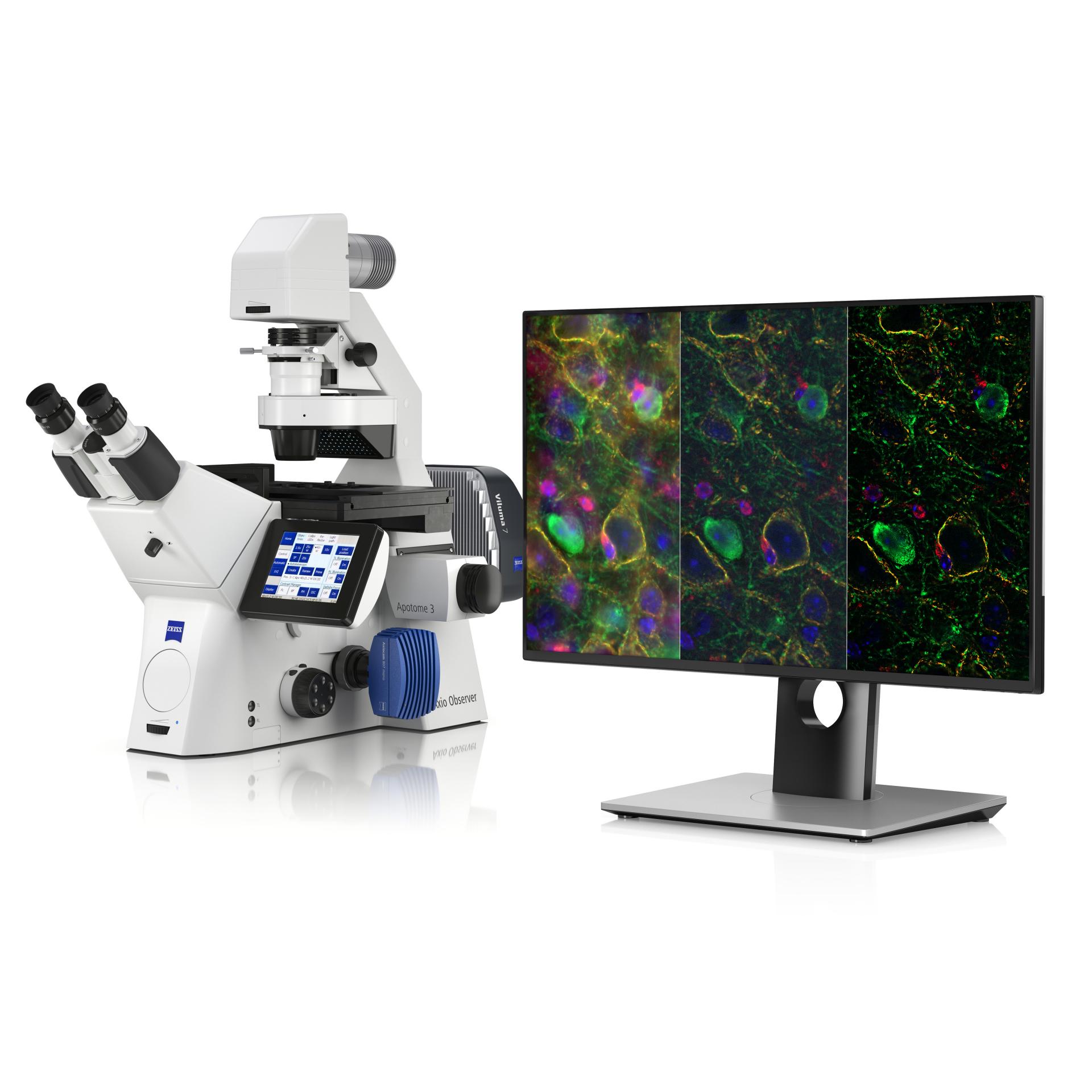

Reveal Hidden Structures with Apotome Plus
Sharper images. Greater insights. More confidence in your data.Apotome Plus enhances ZEISS Apotome 3, bringing confocal-like quality to your widefield microscope. Structured illumination eliminates out-of-focus light, delivering crisp optical sections with lateral resolution down to 180 nm. See deeper into your samples—without the complexity of laser scanning confocal systems.

Why Choose Apotome Plus?
- Unmatched Optical Sectioning – Remove blur and background noise for precise imaging.
- Confocal-Like Clarity – Achieve diffraction-limited lateral resolution in fluorescence microscopy.
- Smart Image Processing with Direct Processing – Process data in real-time, boosting efficiency.
- Reliable, Trustworthy Data – Hardware-based sectioning minimizes artifacts.
- Seamless Integration – Compatible with ZEISS Axio Observer, Axio Imager, and Axio Zoom.V16.


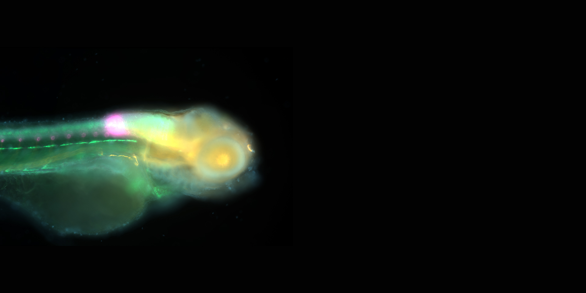
See the Difference
Compare Widefield vs. Apotome vs. Apotome Plus to experience next-level fluorescence imaging.Work Faster with Direct Processing
No waiting. No delays. Just results.Apotome Plus doesn’t just enhance imaging—it makes workflows more efficient. With Direct Processing, image acquisition and data processing happen in parallel, saving time while ensuring optimal sectioning.
✔️ Real-time image processing – Process one dataset while acquiring the next.
✔️ Lossless data compression – Reduce file sizes without compromising quality.
✔️ Seamless workflow integration – Automated processing within ZEISS ZEN software.


How Does It Work?
Apotome Plus refines ZEISS Apotome 3’s structured illumination technology by intelligently processing grid-based images. The result? Sharper optical sections, an improved signal-to-noise ratio, and deeper insights into your samples.
🟢 No lasers.
🟢 No complex alignment.
🟢 Just brilliant images.
Discover the Science Behind Structured Illumination
Access a collection of foundational research articles that explain the principles and advancements of structured illumination microscopy.


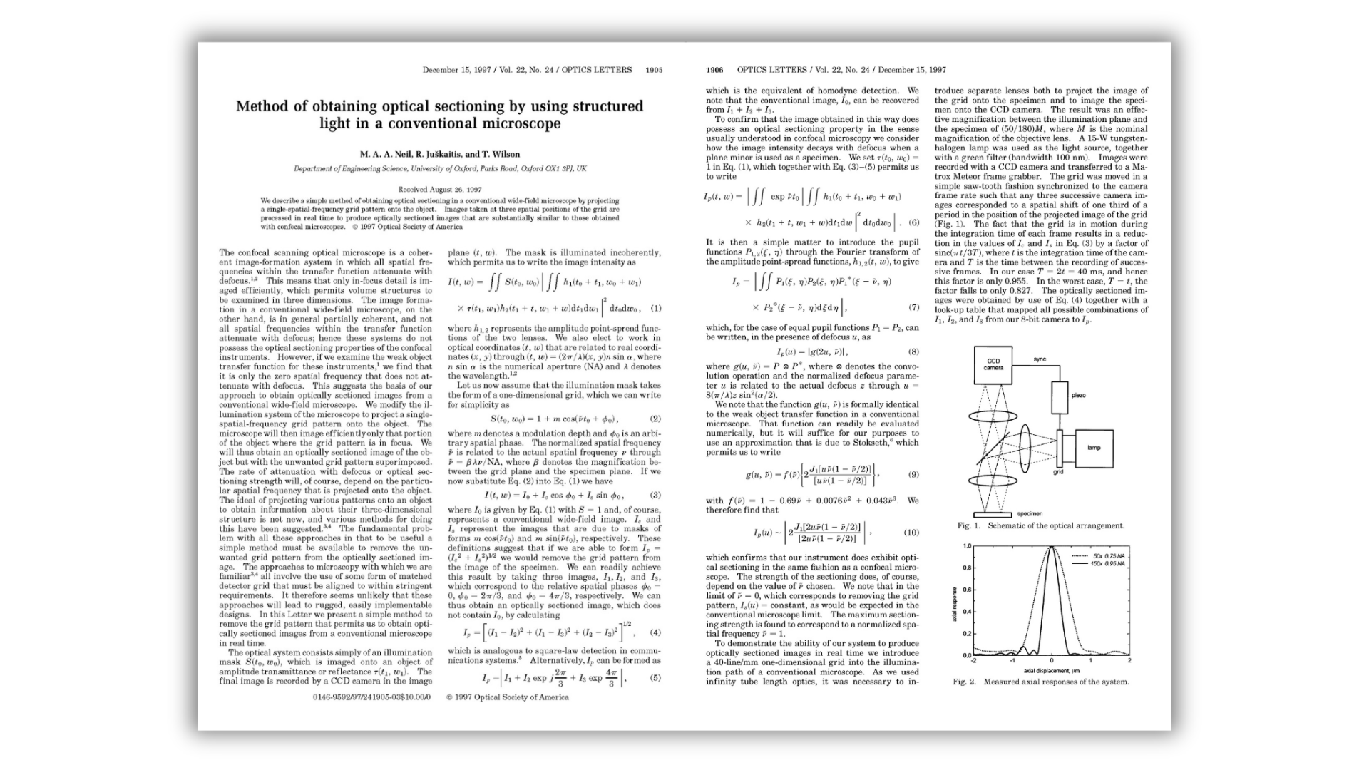

Discover the Science Behind Structured Illumination
Access a collection of foundational research articles that explain the principles and advancements of structured illumination microscopy.What’s Inside?
- The Evolution of Structured Illumination in Fluorescence Microscopy – Explore how structured illumination microscopy (SIM) has revolutionized fluorescence imaging.
- Artifact Reduction and Signal Optimization in Structured Illumination Microscopy – Learn about cutting-edge techniques for maximizing image quality while minimizing distortions.
- The Original Apotome Study: Optical Sectioning Through Structured Light – The research that set the foundation for Apotome technology.
Fill out the form to download your copies.