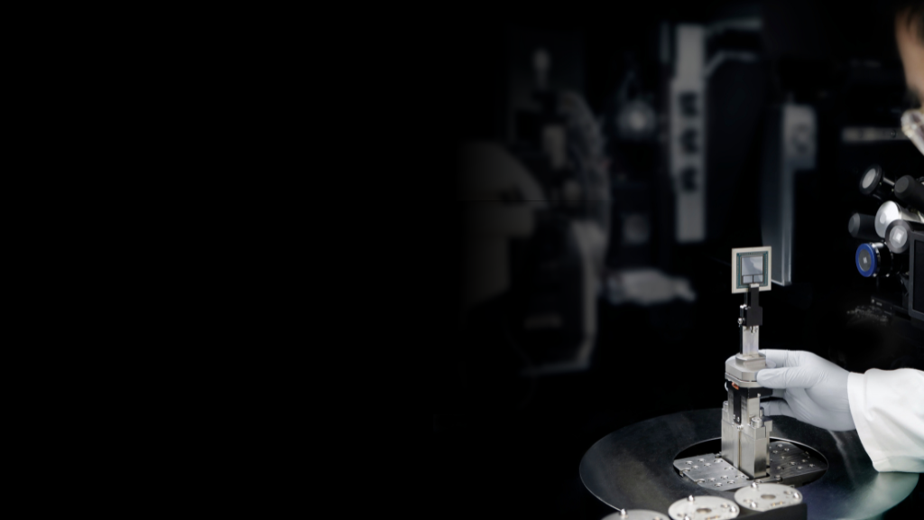

當Deep Learning遇見3D X-ray顯微鏡
ZEISS Xradia 630 Versa 3D X-Ray顯微鏡使用突破性的解析技術,擁有解鎖全新應用的能力,搭載AI智能的新軟體計算邏輯,打破解析度對視野的依賴性,提供更高的效率及更短的分析時間,歡迎參加本次研討會,了解蔡司 X-Ray顯微鏡 (XRM) 在技術上的推進和在材料.電子產品及半導體樣品上的應用。
Breakthrough Imaging with ZEISS Xradia 630 Versa
View highlights of ZEISS Xradia Versa 3D X-ray microscopes: non-destructive imaging, higher resolution with higher throughput.本次線上講座將在Webex平台舉行,您將學習到:
1. 3D X-ray Imaging Systems: 了解3D X-ray 顯微鏡的基礎
2. 了解如何在長工作距離仍可獲得清晰影像
3. 非破壞性的微結構檢測如何幫助您的工作
4. 運用 Advanced Reconstruction Toolbox 3.0 (ART 3.0) 提高您3D X-ray顯微鏡的影像效率
5. 減少金屬偽影與假影修正,提高影像品質
6. 如何運用重構技術在材料科學,電子元件,生命科學研究領域


Head of objectives with the 40X-P
Breakthrough Resolution Performance to Expand Your Research Horizons
The ZEISS 40X-Prime ObjectiveWith more X-ray photons available on ZEISS Xradia 600-series Versa, you can now achieve even faster time to results for varied samples without compromising resolution. Unique to ZEISS Xradia 630 Versa is the 40X-Prime (40X-P) objective lens.
ZEISS Xradia 630 Versa XRM, with the higher energy capabilities of the exclusive 40X-Prime (40X-P) objective, enables you to push the limits of submicron imaging like never before. Known for their ability to achieve resolution at a Distance (RaaD™), ZEISS Xradia Versa platforms allow high resolution imaging of a wide array of sample types and sizes over a long range of length scales.
With 40X-P, the system achieves unparalleled resolution performance of 450-500 nm across the full range of source voltage, from 30 kV to 160 kV, defining RaaD 2.0. Unlocking entirely new application capabilities for researchers, the ZEISS 40X-P objective enables ZEISS Xradia 630 Versa to push industry standards of submicron imaging resolution.
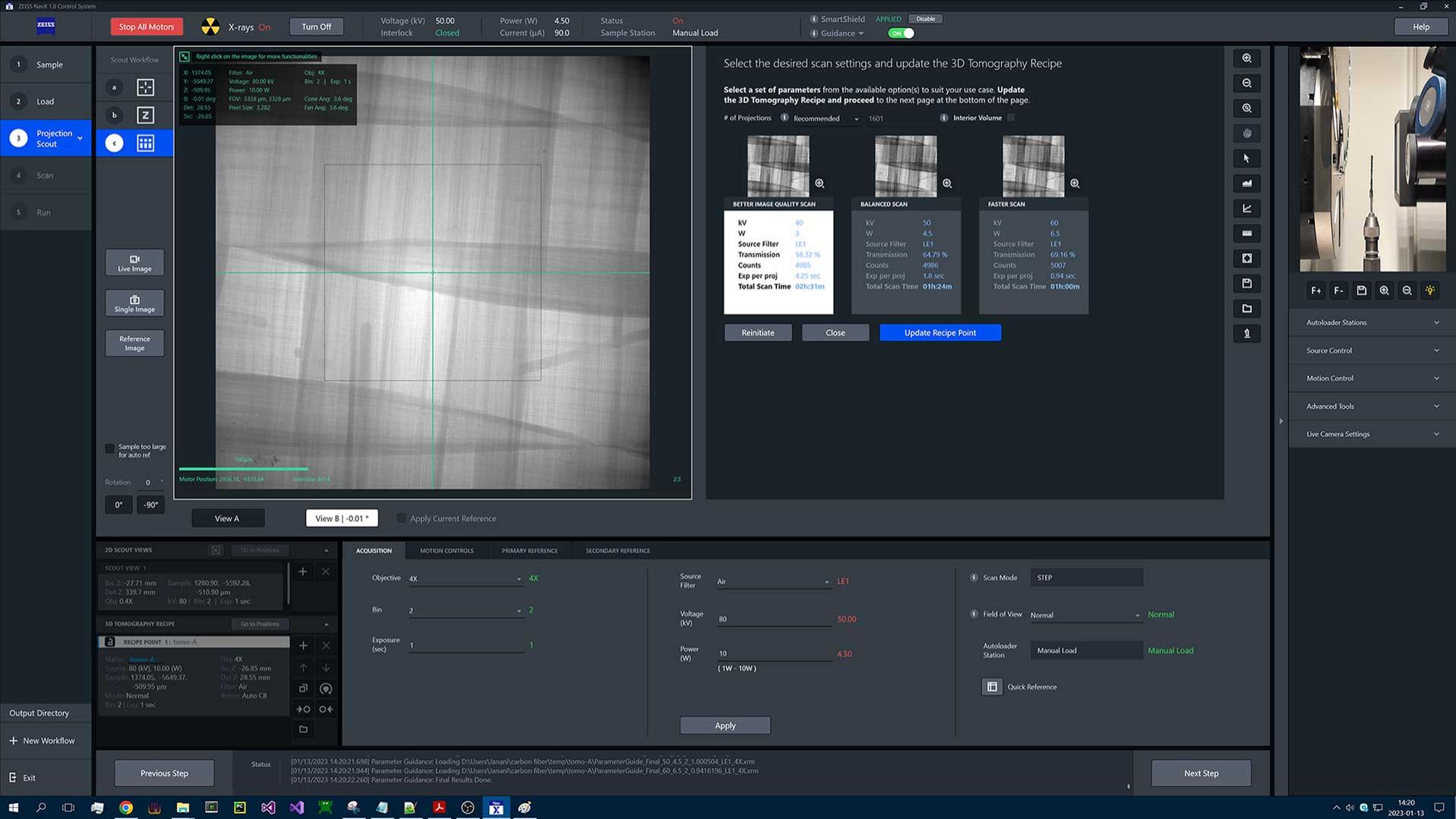

NavX User Interface
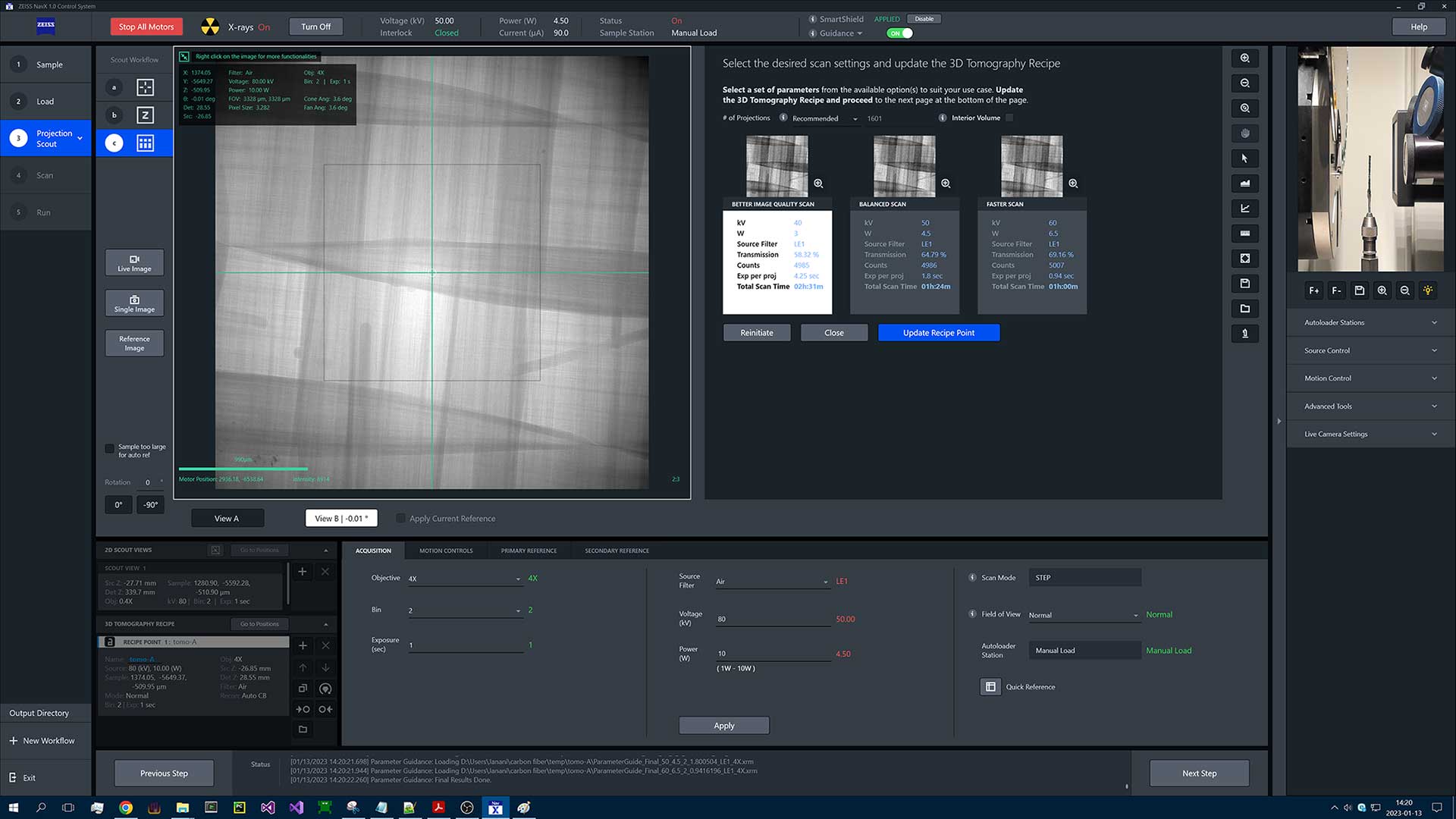

NavX User Interface
NavX User Interface
NavX User Interface
The physics of X-ray imaging can be complex, so ZEISS XRM researchers studied user habits, dove into their challenges, and employed human-centered design (HCD) principles to enable even the newest user in a busy environment to be immediately productive. NavX™, the new user interface for ZEISS Xradia 630 Versa, guides users through automated workflows with intelligent system insights and delivers experimental results more easily and efficiently while also allowing experienced users to explore the full versatility of the platform.
NavX enables you to automate your workflow and provides guidance on the impact the parameters you've chosen will have on your setup. That guidance is directly embedded in the software, taking you through choices in a natural and familiar way.
Additionally, the NavX File Transfer Utility (FTU) takes the data that is being produced by the microscope and automatically transfers it to other locations so that users have their data where they need it, when they need it. These advancements make NavX much more capable for remote operation, advancing user productivity.
NavX intuitive navigation follows the evolution of the XRM user base and revolutionizes X-ray navigation and control with seamless and integrated workflows complementing the planning and execution of advanced correlative workflows..


Flat Panel Extension
The flat panel extension (FPX), standard on your ZEISS Xradia 630 Versa X-ray microscope, further increases the versatility of the system, directly supporting the Advanced Reconstruction Toolbox’s AI-based DeepScout for deep learning and neural network training. Leverage FPX to perform a low resolution, large field of view, "scout” scans, and identify interior regions for higher resolution “zoom” scans on a variety of different sample types. The Volume Scout workflow streamlines this process within NavX.
Advanced Reconstruction Toolbox (ART)
讓您的 ZEISS Xradia X-ray 顯微鏡如虎添翼
蔡司突破性地整合人工智慧,推出最新的ZEISS Advanced Reconstruction Toolbox (ART) 3.0軟體。
憑藉豐富的研究經驗,以及各個領域包含材料科學,半導體,電子材料和生命科學累積的應用實力,發展出嶄新的演算法,使您能更快速、更有效率的取得更清晰的影像結果。
ZEISS Advanced Reconstruction Toolbox (ART) 3.0軟體模組有
- DeepScout software
- DeepRecon Pro
- Material Aware Reconstruction Solution (MARS)
- PhaseEvolve
- OptiRecon
An A12 smartphone package acquired using DeepScout software
For a large field of view scan, a high res scan to train the model, and high resolution reconstruction to LFOV.


ZEISS DeepScout software
Resolution at field of view, throughput at field of viewZEISS DeepScout uses high-resolution 3D microscopy datasets as training data for lower resolution, larger field of view datasets and upscales the larger volume data using a neural network model. ZEISS DeepScout, developed through continued algorithmic innovation enabled by the AI infrastructure from ZEISS, employs the unique Scout-and-Zoom capability to acquire richer information at higher resolution, including interior tomographies for large samples.
- Take your large overview scan
- Feed it through the ZEISS DeepScout reconstruction algorithm
- Get resolution that approaches the resolution of a Zoom scan, but over a much larger field of view.
At its core, ZEISS DeepScout relies on the ability to generate multiscale, spatially registered datasets and uses that ability to train neural networks to improve the reconstruction. New capabilities, fueled by deep learning, mitigate the traditional trade-off between field of view and resolution.
DeepScout, on the left, shows significantly more cellular information than standard reconstruction, on the right.
Sample courtesy of Keith Duncan, Donal Danforth Plant Science Center.
DeepScout
How it worksPolymer electrolyte fuel cell (PEFC) Now, your volume scout includes the full field of view for your sample. A selected high-resolution scan trains the whole model to provide you with high resolution at FOV! This is game-changing AI, uniquely enabling visualization of fine structure across large fields of view at unprecedented speeds.


ZEISS DeepRecon Pro
Harvest the hidden opportunities in big data generated by your XRMThe first commercially available deep learning reconstruction technology enables you to increase throughput by up to 10× without sacrificing novel resolution at a distance (RaaD). Alternatively, keep the same number of projections and enhance the image quality further. ZEISS DeepRecon provides significant AI-driven speed or image quality improvement.
ZEISS DeepRecon Pro is applicable to both unique samples as well as semi-repetitive and repetitive workflows. Self-train new machine learning network models on-site with an extremely easy-to-use interface. The one-click workflow of ZEISS DeepRecon Pro eliminates the need for a machine learning expert and can be seamlessly operated by even a novice user.
ZEISS DeepRecon Pro is now available on ZEISS Xradia Ultra nanoscale XRM.
Mouse lung imaged with Xradia Versa. Sample is iodine stained and captured with 3001 projections. Reconstruction done using DeepRecon (right). Compared with the equivalent image reconstructed using FDK (left)


DeepRecon Pro
ExamplefcBGA flip chip imaged with ZEISS Xradia UltraXRM
left: Standard reconstruction, 1000 projections, 18-hour scan
right: DeepRecon Pro Ultra reconstruction, 250 projections, 4.5 hour scan, a 4x improvement.


Materials Aware Reconstruction Solution (MARS)
Superior image quality for highly attenuating samplesMARS is a reconstruction algorithm that is aware of the constituents within a reconstruction. A challenge in X-ray reconstruction in a lab setting is that imaging with a polychromatic source creates different X-ray energies to generate a phenomenon called beam hardening. This effect is particularly challenging when your material is very dense and embedded in relatively less dense material. MARS tells the reconstruction system how to compensate for the effect of extreme beam hardening in the regions between very dense objects. This is important in applications like biomaterials, where you might be looking at implants next to bone or tissue. Or electronics where extremely dense solder balls appear next to other less dense materials on a printed circuit board, generating strong artifacts. MARS reconstructs your images to compensate for these effects.
Biomedical metal implant in bone. Without MARS, left. With MARS, right.


PhaseEvolve
Enhanced image contrast and improved segmentationZEISS PhaseEvolve is a patent-pending postprocessing reconstruction algorithm that enhances the image contrast by revealing material contrast uniquely inherent to X-ray microscopy, which can often be obscured by phase effects in low-medium density samples or high-resolution datasets. Perform more accurate quantitative analysis with improved contrast and segmentation of your results.
Rayon fibers were imaged at 1.5 μm/voxel resolution and processed using ZEISS PhaseEvolve revealing the large distribution of radial porosity along the length of the fibers.
Sample courtesy Dr. Sherry Mayo & Dr. David Fox, CSIRO, Australia.


ZEISS OptiRecon
Fast and efficient iterative reconstruction solutionA fast and efficient algorithm-based technology that delivers iterative reconstruction from your desktop, allowing you to achieve up to 4× faster scan times or enhanced image quality with equivalent throughput. ZEISS OptiRecon is an economical solution offering superior interior tomography or throughput on a broad class of samples.
Camera module: 1200 projections in 90 minutes using standard FDK, left, vs. 300 projections in 22 minutes with OptiRecon, right. Comparable image quality in a fraction of the time.
PhaseEvolve
Enhanced image contrast and improved segmentationZEISS PhaseEvolve is a patent-pending postprocessing reconstruction algorithm that enhances the image contrast by revealing material contrast uniquely inherent to X-ray microscopy, which can often be obscured by phase effects in low-medium density samples or high-resolution datasets. Perform more accurate quantitative analysis with improved contrast and segmentation of your results.
Rayon fibers were imaged at 1.5 μm/voxel resolution and processed using ZEISS PhaseEvolve revealing the large distribution of radial porosity along the length of the fibers.
Sample courtesy Dr. Sherry Mayo & Dr. David Fox, CSIRO, Australia.
當Deep Learning遇見3D X-ray顯微鏡
線上講座- 3D X-ray Imaging Systems: 了解3D X-ray 顯微鏡的基礎
- 了解如何在長工作距離仍可獲得清晰影像
- 非破壞性的微結構檢測如何幫助您的工作
- 運用 Advanced Reconstruction Toolbox 3.0 (ART 3.0) 提高您3D X-ray顯微鏡的影像效率
- 減少金屬偽影與假影修正,提高影像品質
- 如何運用重構技術在材料科學,電子元件,生命科學研究領域


Nicky Liu is responsible for applications on ZEISS X-ray microscope (XRM) product portfolio and familiar with Scanning Electron Microscope (SEM). With experienced consultative skills, she can investigate the true problems that customer may have and suggest best usage scenario and product adaptation. Her past experiences working in National Cheng Kung University and Bruker AXS enables her capability to address academic researcher’s and semiconductor industry customers' pain points. Nicky is also the focal point to provide feedback to ZEISS Product Development Team.